

Chicago, IL 60611 United States


Overview
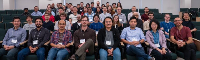
Advances in high-throughput imaging techniques now generate massive amounts of multiscale image data, ranging from whole tissues to single cells to protein structures. Beyond static snapshots, novel imaging methods capture long time-series that provide valuable spatio-temporal information. Analyzing this unprecedented volume of biological data raises a variety of mathematical challenges. Notably, how can we model biological variation while disentangling biological variation from technical heterogeneity? For example, how do single cells grow and progress through the cell cycle under diverse stimuli, or how do proteins and molecular complexes undergo conformational changes? Despite the richness of these data, methods for spatio-temporal analysis remain limited in computational biology. At the same time, tools from multiple disciplines—such as deep learning in computer science, mechanistic models in biophysics, and dynamical systems theory in mathematics—have been developed to study related problems. In this context, our proposed workshop will focus on theoretical and computational tools from mathematics and physics that enable rigorous analysis of spatio-temporal biological data, with the goal of elucidating the temporal dynamics of underlying biological processes.
Intended for a diverse audience of mathematicians, physicists, and computational and experimental biologists, the workshop will feature complimentary sessions on different facets of imaging data, with a particular focus on dynamics. Our interdisciplinary organizing team will begin with a general overview of shape space, highlighting both theoretical foundations and state-of-the-art computational tools. We will then present several publicly available datasets on protein and cell shapes from advanced imaging experiments, and brainstorm methods to uncover the dynamics underlying observed heterogeneity. The ultimate goal of the workshop is to develop a blueprint for new frameworks to study dynamics in shape space and to foster collaborations across mathematics, computer science, and biology.
Schedule
October 19 2026 Monday
8:30 am - 8:55 am
Breakfast
9:00 am - 9:15 am
Welcome & announcements - NITMB director
9:15 am - 9:30 am
Welcome from organizers and overview of the workshop structure
9:30 am - 11:40 am
Session1: Biophysics: Mechanics and Physical Models of Cell Shape
Chair: Ashok Prasad
9:30 am - 9:55 am
Mattia Serra
Low-dimensional representations of tissue flows for quantifying and controlling morphogenesis
During embryonic development, thousands of cells move collectively to generate reproducible tissue flows and dynamic embryo morphology. How can we represent/compress these spatiotemporal flows to support quantitative comparison, mechanistic understanding, and control? I will present a dynamical systems framework that identifies coherent structures—attractors and repellers of cell trajectories—from imaging data. Combined with physical models and experimental perturbations, these representations have revealed new mechanisms controlling avian gastrulation flows and embryo geometry, organizing distinct phenotypes into a phase diagram with biologically interpretable axes. Finally, I will present ongoing work using equivariant variational autoencoders to learn low-dimensional representations of morphogenetic flows to quantify variability within and across phenotypes.
9:55 am - 10:20 am
Min Wu
Coupling Shape in Physical Space and State Space
Cellular organization has shape in two spaces: physical space, defined by the morphology of cells and organelles, and state space, where molecular activities evolve on geometrical structures defined by the underlying dynamical system. I will introduce our experimental work suggesting that these two forms of geometry are coupled, using examples of cortical signaling waves that interact with the membrane bilayer and communicate with organelle morphology. I will also discuss our efforts to identify the dynamical variables and low-dimensional network that link trajectories in molecular state space to evolving cell shapes in physical space (e.g. cell division), with the broader goal of understanding physical shape and dynamical state as coupled representations of cellular organization.
10:20 am - 10:50 am
Coffee Break
10:50 am - 11:15 am
Trang Le
Cellular Morphology and Spatial Proteome Organization
How does cellular shape relate to the spatial organization of the proteome? The Shapespace framework combines cellular shape analysis, spatial proteomics, and machine learning to explore this relationship. Analyzing over one million cells spanning 11,998 proteins across 11 human cell lines, we identified a shared continuum of cell and nuclear shapes. Organelle topology remained stable across shape space within each cell line but differed between cell lines, while individual proteins exhibited shape-associated abundance variation, including proteins unrelated to the cell cycle. A common spatial coordinate framework enables comparison of protein distributions across shapes and under drug perturbation. Together with complementary models predict single-cell protein localization and generate virtual organelle staining from microscopy images, these approaches connect morphological variation with molecular organization, providing a foundation for studying cellular states and dynamics—and a step toward a data-driven model of the cell.
11:15 am - 11:40 am
Yogesh Goyal
From Cells to Systems: Learning the Spatiotemporal Rules of Aortic Growth
11:40 am - 1:00 pm
Lunch
1:00 pm - 2:40 pm
Session 2 - Imaging and Experimental Cell Biology
Chair: Matheus Viana
1:00 pm - 1:25 pm
Brandon Schlomann
Deciphering Gene Expression Patterns in Dynamic Tissues
Living tissues generate intricate spatial patterns of gene expression that are essential to their function; being able to quantitatively interpret these patterns would help identify important underlying cellular processes. I will discuss our work using live imaging of transcriptional dynamics to decipher such patterns in two model systems: coupled genetic oscillators in zebrafish development and immune response to bacterial infection in fruit flies. These case studies indicate that, even in simple examples, interpreting spatial patterns of gene expression is practically impossible without direct measurement of dynamics and their response to perturbations. I will conclude by describing our ongoing attempts to formulate and parameterize stochastic models of collective gene expression at the tissue scale, where efficiently incorporating realistic tissue geometry remains an open challenge
1:25 pm - 1:50 pm
Alex Khang
Investigating Single Cell Shape and Dynamics Within hIPSC-derived Lumenoids
Tissue morphology and cell shape are intimately linked but how this relationship is established, and how it changes over time, remains poorly understood. We study this problem using hiPSC-derived lumenoids which are spherical self-organizing stem-cell structures that provide a tractable model for studying early epithelial organization. We show that individual cell shape varies systematically with position within the lumenoid and with lumenoid size, revealing a coupling between local cell geometry and global tissue architecture. Using 5D timelapse imaging, we also track how cells move collectively and how lumenoid shape changes over time, demonstrating that this is not just a multi-scale problem but also a dynamic one operating across multiple timescales. We are currently extending this work to examine cell-cell interfaces and vertex model simulations, with the aim of building a multiscale understanding of how cell and tissue properties are coupled. This talk will highlight open questions around cell positional information, mechanical feedback, and the rules governing cell packing and organization.
1:50 pm - 2:15 pm
Aaron van Loon
Data-driven Discovery of Morphodynamic Cycles During 3D Neutrophil Motility
Neutrophils rapidly change their morphology as they navigate complex interstitial environments to reach sites of inflammation and infection. To interrogate the relationship between morphological change and motility in 3D, we observed membrane-labeled dHL-60 neutrophils spontaneously migrating through collagen hydrogels using high spatial and temporal resolution. Prior to 3D shape analysis, we considered three different alignment frames which would encode different shape and behavior information into the shape modes we extract from principal component analysis on spherical harmonic coefficients from segmented cell surfaces. The “shape-only” frame aligns to the first two principal axes of the cell volume coordinates, the “trajectory + shape” frame aligns to the cell’s trajectory vector and the vector of the greatest mass orthogonal to the trajectory, and the “trajectory-only” frame aligns to the cell’s current and next trajectory vectors. The top principal components (PCs) from each of these alignments represent similar morphological features including cell orientation, aspect ratio, and volume distribution, but the interpretation of PCs across these shape spaces differs depending on how each PC relates to its respective alignment frame. To quantify morphological dynamics, we calculate the magnitude and direction of transitions between discrete states in coarse-grained phase spaces (CGPSs) defined by pairs of the top PCs for each alignment frame. Morphodynamics across CGPSs generally increased with the number of trajectory axes used for alignment; interestingly, flux in many of these phase spaces appeared cyclical. The CGPS defined by PC1 and PC2 in the trajectory-only frame had by far the highest cyclical activity as measured by area enclosing rate and cycle period. By observing the activity of individual cells in this CGPS for up to an hour, we found that cells dwell in a region of the space associated with elongation along their trajectory while moving persistently in real space, then perform a circuit in the CGPS correlated with rapid rear retraction and directional change in real space. This cycle quantifies a well-established neutrophil behavior during spontaneous motility, in which persistent motility is periodically interrupted by a pause during which a new motility direction is chosen.
2:15 pm - 2:40 pm
Francis Lin/Nick Palmerley
Long-lasting Giant Blebbing of Human Breast Cancer Cells Induced by Microfluidic Confinement
How individual cancer cells adapt their morphology to confinement, and how these responses are influenced by the mechanical stiffness and chemical cues of the surrounding microenvironment, remains poorly understood. One prominent cellular response is membrane blebbing, characterized by the formation of balloon-like protrusions of the cell membrane. This raises the fundamental question of how confinement dimensionality, mechanical stiffness, and chemical cues regulate bleb formation and dynamics during cancer invasion. To address this question, we developed an integrated microfluidic platform that combines three-dimensional confinement, controlled mechanical stiffness through variation of polydimethylsiloxane (PDMS) properties, and chemical gradients across hundreds of cell-trapping compartments. Using MDA-MB-231 cells as a TNBC model, together with live-cell imaging and quantitative morphology analysis, we investigated how these physical and chemical conditions influence blebbing and cellular protrusion behavior. Comparisons across confinement dimensions and stiffnesses provided a means to examine how mechanical properties shape bleb dynamics. Lastly, these observations provided the basis for a conceptual biophysical framework describing how confinement dimensionality and stiffness regulate bleb formation. Altogether, this work provides a quantitative approach for studying how cancer cells adapt their morphology to mechanically heterogeneous tissue environments and offers new insight into the biophysical regulation of membrane blebbing during cancer invasion.
2:40 pm - 3:10 pm
Coffee Break
3:10 pm - 4:00 pm
Tutorial, Part 1 - Wenjun Zhao, Khanh Dao Duc, Shira Faigenbaum-Golovin
4:00 pm - 5:00 pm
Tutorial, Part 2 - Wenjun Zhao, Khanh Dao Duc, Shira Faigenbaum-Golovin
October 20 2026 Tuesday
8:30 am - 8:55 am
Breakfast
9:00 am - 9:30 am
Gathering in the Auditorium
9:30 am - 11:40 am
Session 3 - Theory: Mathematical Foundations for Shape Analysis
Chairs: Khanh Dao Duc, Shira Faigenbaum-Golovin, Wenjun Zhao
9:30 am - 9:55 am
Dominic Skinner
Quantifying Variation Along Morphological Trajectories in Drosophila Larval Trachea
Cells, tissues, and organs, undergo dramatic morphological transformations during development. Increasingly it is experimentally accessible to measure measure three dimensional morphology across time, yet it remains challenging to use these measurements to systematically analyze trajectories such as by finding average trajectories and quantifying variability. Here, we study the Drosophila larval trachea, where dorsal branches undergo extreme morphological changes forming complex and highly non-convex shapes. By representing diverse branch shapes in a common space, we are able to identify directions of morphological variation and directions along which successful and unsuccessfully differentiated branches vary along. Through regularized dynamic time warping, we can compare entire trajectories, finding a trajectory barycenter, as well as demonstrating that successful branches in a genetically perturbed system are delayed relative to successful branches in the unperturbed system.
9:55 am - 10:20 am
Laurent Younes
Shape Alignment via Allen-Cahn Nonlinear-Convection
We describe a phase-field method for shape registration to align shapes of possibly different topology. A soft end-point optimal control problem is introduced whose cost function measures the control norm required to align an initial shape to a final shape, up to a small error term. The initial data is spatially integrable, the paths in control spaces are integrable and the evolution equation is a generalized convective Allen-Cahn. Inspired by level-set methods and large diffeomorphic deformation metric mapping, the controls spaces are integrable scalar functions to serve as a normal velocity and smooth reproducing kernel Hilbert spaces to serve as velocity vector fields. The existence of mild solutions to the evolution equation is proved, and numerical simulations of minimums to the fully discretized optimal control problem are displayed. This research offers a novel discrepancy measure that provides valuable ways to analyze diverse image data sets. This is joint work with Daniel Solano and Jérôme Darbon, completed during Daniel Solano’s Ph.D. thesis.
10:20 am - 10:50 am
Coffee Break
10:50 am - 11:15 am
Anuj Srivastava
Statistical Shape Analysis of Complex Biological Structures
Advances in imaging technologies have made quantitative morphometrics increasingly important for understanding biological form and function. This introductory talk presents a statistical framework for analyzing complex biological objects — including cells, chromosomes, brain structures, plants, and vascular networks — represented as curves, surfaces, trees, and graphs. Elastic Riemannian metrics provide principled comparisons that are invariant to rotation, translation, scale, and parameterization, enabling statistical analyses such as estimation of mean shapes, principal component analysis of shape variation, population modeling, and analysis of temporal shape dynamics. By characterizing both cross-sectional variability and longitudinal evolution, this framework supports the study of morphometrics and morphogenesis across diverse applications, including relating mitochondrial morphology to lifestyle, constructing shape alphabets for symbolic representations of the three-dimensional genome, predicting cognitive decline from brain anatomy, and quantifying age-related changes in cerebral arterial networks.
11:15 am - 11:40 am
Zixuan Cang
Optimal Transport Across Biological Representations
11:40 am - 1:00 pm
Lunch
1:00 pm - 2:40 pm
Session 4 - Data-Driven Analysis: Image Analysis, Machine Learning and Inference
Chair: Felix Zhou
1:00 pm - 1:25 pm
Keisuke Ishihara
An Integral Geometry Approach to 3D Biological Shape Analysis with Minkowski Tensors
The biomedical sciences is witnessing an explosion of volumetric imaging data. Morphology offers a quantitative readout of cell state and organ pathology, yet extracting meaningful shape features from segmented 3D structures remains a bottleneck. Widely used descriptors such as CellProfiler and spherical harmonic descriptors lack principled rotational invariance and scale poorly with geometric complexity, limiting their utility for systematic morphological phenotyping. We propose Minkowski tensors (MinT) as a physically interpretable 3D shape descriptor. Rooted in integral geometry, MinT jointly encode volume, surface area, curvature, and anisotropy in a compact tensor representation. We introduce pykarambola, an open-source Python package that computes Minkowski tensors directly from segmented 3D label images and meshes. Using a systematic linear classification benchmark across three datasets spanning nuclear morphology and 3D tissue structures, we show that MinT-derived features consistently outperform both CellProfiler and spherical harmonic descriptor feature sets, while using substantially fewer features. MinT achieves classification accuracy higher than ResNet-based image classifiers, without requiring network training. These results establish Minkowski tensors as an expressive, compact, and interpretable foundation for shape-based phenotyping of 3D biological structures. In the future, MinT maybe integrated into generative modeling workflows for 3D shape: as a structured latent space, a regularizer, or an evaluation metric.
1:25 pm - 1:50 pm
Petia M. Vlahovska
Learning Membrane Properties from Shape Fluctuations and Deformation of Cell-Mimetic Vesicles
The dynamic shape of a biological membrane encodes information about its underlying electromechanical properties. We use giant unilamellar vesicles as a cell-mimetic experimental platform in which spontaneous membrane shape fluctuations and electric-field-induced deformation are recorded by optical microscopy and analyzed using continuum-mechanics models. The amplitude and temporal evolution of these shape dynamics provide quantitative measurements of membrane bending rigidity, tension, viscosity, and capacitance. I will discuss how combining time-resolved imaging with mechanistic models allows membrane material properties to be inferred from shape dynamics, as well as the challenges that arise when membrane properties are spatially heterogeneous or the vesicle departs from a spherical shape.
1:50 pm - 2:15 pm
Chuangqi Wang
From 2D to 3D: Resolving Heterogeneity in Subcellular Protrusion Dynamics
Protrusion dynamics reflect coordinated cytoskeletal regulation that shapes cell migration, antigen presentation, cell-cell interactions, and beyond. Revealing that coordination from live-cell imaging is fundamentally a computational problem: extracting meaningful representations from raw time-series data in 2D – a challenge that grows substantially harder in 3D. During my PhD, monitoring the PtK1 epithelial cell line on the 2D substrates, utilizing the conventional Machine Learning and Deep Learning techniques, we developed HACKS/DeepHACX frameworks that represents local shape dynamics as edge-velocity time series sampled within small probing windows, and learns interpretable, perturbation-sensitive features. This representation resolved fine-grained protrusion phenotypes and linked them to specific actin regulators coordination, including Arp2/3 and VASP. Since starting my independent lab, I have begun extending this representation into three dimensions, applying local surface-based sampling to live 3D imaging of primary T cells from mice in the in-vitro 3D gel model in collaboration with Dr. Jordan Jacobelli. Unlike adherent epithelial cells, T cells are smaller and far more dynamic, with no stable 2D boundary to window: their protrusions form and retract rapidly across a deforming 3D surface, requiring a fundamentally different representation of shape dynamics than a fixed edge in a plane. Building on the Uwrap3D framework for 3D surface reconstruction, collaborated with the Felix Zhu Lab, I will discuss the computational challenges of representing and learning shape dynamics on non-adherent, volumetric geometries, and the promise this approach holds for linking actin regulator function to physiologically relevant protrusion heterogeneity in 3D cell migration.
2:15 pm - 2:40 pm
Tzer Han Tan
Mapping Developmental Shape Space through Symmetry and Representation Learning
Stem-cell-based embryos and organoids are powerful models of development, but their substantial morphological variability makes it difficult to define reproducible developmental states and trajectories. We ask whether this variability can be organized into a low-dimensional and biologically interpretable landscape of shape. To do so, we combine variational autoencoders with symmetry-based spectral decompositions, including Zernike modes, to identify interpretable morphological features and broken symmetries. Applied to synthetic embryo datasets, this framework captures key transitions, including anterior-posterior polarization and elongation. Correlating latent coordinates with Zernike modes links learned dimensions to specific symmetry-breaking processes. Projecting developmental trajectories into low dimensions further reveals effective morphological flow fields that organize how embryos move through shape space. Most excitingly, the generative capacity of the VAE allows us to reconstruct virtual developmental trajectories toward target morphologies, creating a potential route from learned representations to experimentally testable predictions.
2:40 pm - 3:10 pm
Coffee Break
3:10 pm - 4:00 pm
Questions/Data - Liya Ding and Matheus Viana
4:00 pm - 5:00 pm
Biologists present data and problems
5:00 - 6:00 pm
Reception (counts as dinner)
October 21 2026 Wednesday
8:30 am - 8:55 am
Breakfast
9:00 am
Gathering in the Auditorium
9:30 - 10:20 am
Housekeeping and Breakout Sessions
10:20 - 10:50 am
Coffee Break
11:00 am - 11:40 am
Breakouts
11:40 pm - 1:00 pm
Lunch
1:00 - 2:40 pm
Breakout sessions
2:40 pm - 3:10 pm
Coffee Break
3:10 pm - 4:00 pm
Breakout reports
4:00 pm - 5:00 pm
Poster Session
October 22 2026 Thursday
8:30 am - 9:00 am
Breakfast
9:00 am - 9:15 am
Gathering in the Auditorium
9:15 am - 10:00 am
Toward a Research Program for Representing and Learning Morphology & Shape Dynamics for Biological Data
10:00 am - 10:15 am
Coffee Break
10:15 am - 11:00 am
Breakouts
11:00 am - 12:00 pm
Breakouts
12:00 - 1:00 pm
Lunch
1:00 - 2:00 pm
Breakouts
2:00 - 3:00 pm
Breakouts
3:00 - 3:15 pm
Coffee Break
3:15 - 4:00 pm
Breakout Reports
4:00 - 5:00 pm
Breakout Reports
October 23 2026 Friday
8:30 - 9:00 am
Breakfast
9:00 - 9:15 am
Gathering in the Auditorium
9:15 - 10:00 am
Toward a Research Program for Representing and Learning Morphology & Shape Dynamics for Biological Data
10:00 - 11:00 am
NITMB Seminar - Julie Theriot - Cells Getting In Shape For Rapid Directed Migration
Rapid actin-based crawling of eukaryotic cells depends on coordination of multiple dynamic cellular elements over a wide range of distance and time scales. Persistent, rapid directional migration involves the balanced interaction of forces generated by actin filament assembly and disassembly, myosin-based network contraction and demolition, cell-substrate adhesion, membrane tension, and hydrostatic pressure within the cell. This same set of subcellular structures, forces and interactions also determines cell shape; indeed, for many cell types the direction of movement and approximate instantaneous speed can be estimated with reasonable accuracy from static snapshots. We have used quantitative videomicroscopy as a primary tool to explore mechanisms determining the speed and persistence of actin-based cell motility, as well as origins of cell-to-cell variation, in a variety of motile systems that operate in 1D, 2D and 3D spaces. We focus on the fastest-moving animal cells including fish epidermal keratocytes and mammalian neutrophils, with typical speeds on the order of 10-20 µm/min, or about 100 times faster than most fibroblasts or immortalized cell ines. We have developed and optimized a wide range of methods for extraction of interpretable quantitative measurements of cell shape and dynamics from videomicroscopy data, with the goal of maintaining an accurate description of biologically important features in the data, while reducing the dimensionality and also minimizing the propagation of experimental noise. Most recently we have applied these methods to discovering stereotyped sequences of 3D shape changes for the movement of human neutrophils navigating in loose collagen matrices.
11:00 am - 12:00 pm
Concluding remarks and free time
12:00 - 1:00 pm
Lunch
1:00 pm
End
Participants
Organizers
- Khanh Dao Duc – University of British Columbia
- Shira Faigenbaum-Golovin – Bar Ilan University
- Matheus Palhares Viana – Allen Institute for Cell Science
- Ashok Prasad – Colorado State University
- Wenjun Zhao – Wake Forest University
- Felix Zhou – Vanderbilt University
Participants
- Ratul Biswas – NITMB Fellow
- Monica Brown – Vanderbilt University
- Zixuan Cang – North Carolina State University
- Rich Carthew – Northwestern University
- Xiaolong “Harry” Chen – University of California, San Diego
- Seo Woo Choi – LifeCanvas Technologies Inc.
- Luca Deininger – ETH Zurich
- Willem Diepeveen – University of California, Los Angeles
- Liya Ding, The University of Chicago
- Alex Eskandarian – Harvard Medical School
- Venkatesh Gopal – Elmhurst University
- Yogesh Goyal – Northwestern University
- Felix Granum – Duke University
- Alasdair Hastewell – NITMB Fellow
- Edwin Huras – University of British Columbia
- Keisuke Ishihara – University of Pittsburgh
- Rubaiyat Bin Islam – Tulane University
- Ece Karacam – Florida State University
- Alex Khang – Allen Institute
- Kazuki Koga – Institute of Science Tokyo
- Yuyang Lai – Department of Physics, University of California San Diego
- Adele Lantow – UC Santa Barbara
- Matt Lastner – University of Utah
- Ngoc Hai Trang Le – Biohub
- Eardi Lila – University of Washington
- Francis Lin – University of Manitoba
- Milton Lin – NITMB Fellow
- Atsushi Matsuda – University of Washington
- Stathis Megas – Medical University of Vienna
- Xiangyi “X” Meng – Rensselaer Polytechnic Institute
- Xuan Ouyang – UC San Diego
- Nick Palmerley – University of Manitoba
- Mariya Savinov – NITMB Fellow
- Brandon Schlomann – Simon Fraser University
- Mattia Serra – UC San Diego
- Ioannis Sgouralis – University of Tennessee Knoxville
- Dominic Skinner – Flatiron Institute
- Anuj Srivastava – Johns Hopkins University
- Tzer Han Tan – University of California San Diego
- Julie Theriot – University of Washington
- Ohad Vilk – NITMB Fellow
- Daniel Tolosa Villarreal – Arizona State University
- Aaron van Loon – University of Washington
- Petia Vlahovska – Northwestern University
- Chuangqi Wang – University of Colorado, Anschutz Medical Campus
- Xiaoqi Wei – North Carolina State University
- Alex Wong – Harvard Medical School
- Min Wu – Yale University
- Yuan Young – New Jersey Institute of Technology
- Laurent Younes – Johns Hopkins University
- Gaohan Yu – University of Pittsburgh
- Jiaqi Zhang – Institute for Genome Sciences, University of Maryland School of Medicine
- Yiwen “Sharon” Zhou – The University of Chicago